Our products
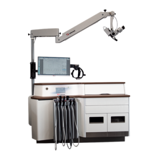
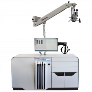
Entermed ENT treatment units are hand built to the highest standards by a team of highly skilled craftsmen. Attention is paid to the smallest detail during the fabrication process resulting in beautifully crafted custom made ENT treatment units and complementary furniture.
-310x310.png)


-260x78.png)
What we stand for
Dedication
Our customers define our products. The ENT treatment units are designed based on their needs.
Innovation
Our products are based on years of innovation. Continuous development defines who we are.
Safety guaranteed
We put safety for doctors and patients first. All of our products are hand built to the highest safety and quality standards.
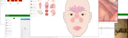
A user-friendly setting
Touch-screen operation, hygienic materials, carefully selected finishes and other elements are combined to form the basis of an efficient, durable and patient-friendly setting.
Don’t let your equipment hinder your performance!
-555x558.png)
We are here to help
Would you like to know more about us or our products, please fill out the form and we will contact you.